Spotlight on ... Cranial Cruciate Ligament Rupture
What is Cranial Cruciate Ligament Rupture?
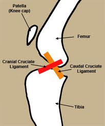
The canine knee (stifle) is a complicated joint. Two major bones meet in this joint, the femur (thigh bone) and the tibia (shin bone). These are joined by the patella (knee cap) and fabellae (very small bones found at the back of the knee). The joint is held together and stabilised by various ligaments. These ligaments make sure that the knee can only bend in ways that it should. There are two cruciate ligaments, the cranial (towards the head) and the caudal (towards the tail). Besides these ligaments there are tendons that attach the bones to muscles and so enable movement of the joint, and menisci which act as shock absorbing cushions.
Within this complicated joint the cranial cruciate ligament has the job of preventing the tibia from moving forward under the femur. When it ruptures, partially or completely, the tibia is able to move forward. This abnormal function results in varying degrees of lameness. The ruptured ligament makes the joint unstable, causes pain, and damage to the joint (for example, tearing of the shock absorbing cushions). If left untreated your dog's lameness may appear to improve in the first week or so, but you will note that the joint looks swollen and lack of treatment simply means that the abnormal movement caused by the ruptured ligament will continue and further degeneration of the joint will occur. In the long term osteoarthritis will develop even in cases that are diagnosed and treated in the acute stages of this disease, but the progression is quicker in chronic cases.
Rarely traumatic, cranial cruciate ligament rupture is more usually the result of a disease process extending over months or years. It is caused by a combination of several factors including degeneration of the ligament, obesity, poor physical condition, conformation, and breed. Many dogs who suffer from cranial cruciate ligament (CCL) rupture in one knee will go on to develop a similar problem in their other knee, and partial tearing of this ligament frequently progresses to a full rupture over time.
What are the signs and symptoms of Cranial Cruciate Ligament Rupture?
The degree of lameness varies from cases to case, but with complete CCL rupture it is likely that you will observe your dog becoming suddenly lame. However, given that many dogs suffer a partial tear and only progress to a full rupture over time it is important to be alert to more subtle signs including your dog's inability to sit square and preference for putting his legs out to the side when sitting down. Other signs to look out for include:
difficulty rising
difficulty jumping into the car
decreased activity level
muscle atrophy (decreased muscle mass in the affected leg)
decreased range of motion of the knee joint
a popping noise (which may indicate a meniscal tear)
swelling on the inside of the joint (fibrosis or scar tissue)
lameness
In chronic cases where the partial tear or rupture occurred some time ago the signs and symptoms will be akin to those seen with osteoarthritis:
decreased activity
stiffness
unwillingness to play
pain
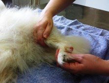
In addition to taking a history from you, your veterinarian will manipulate the joint in order to test whether and to what extent the tibia is able to move forward under the femur. She may also take x-rays to confirm the presence of fluid in the joint, the progression of osteoarthritis, to aid in planning surgical treatment, and to rule out any other conditions that may be present. These x-rays will not enable your veterinarian to establish the extent of damage to the CCL or to the menisci. This can only be established through surgical investigation.
What treatments are available?
Surgical and non-surgical (also known as conservative) treatments are available, and your veterinarian will assist you in determining which is most appropriate for your dog. The most appropriate option for your pet will depend on your pets activity level, size, age, and conformation, the degree of knee instability, and other factors, such as the progression of osteoarthritis.
Surgical treatments are generally preferable when dealing with CCL rupture since they provide a means of permanently addressing the instability of the joint and any damage to the menisci. The surgical options available for controlling the joint's instability are varied and will depend on whether the surgery is carried out by a general practitioner or a specialist.
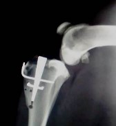
Specialist surgeons may choose to use procedures such as the Tibial Plateau Levelling Osteotomy (TPLO), Tibial Tuberosity Advancement (TTA) or Modified Maquet Procedure (MMP), which seek to address and control the instability caused by the ruptured CCL by changing the biomechanics of the joint. These techniques involve making a cut in the tibia. These procedures have demonstrated high degrees of success in enabling dogs to return to normal function after CCL rupture with a low level of complications.
The surgical techniques which involve cutting the tibia, TPLO, TTA, and MMP, have superior results with regard to limb function and the progression of osteoarthritis compared to traditional suture techniques, and are often favoured in cases involving young, large breed dogs. The disadvantage of such techniques is related to the cut made in the bone which requires time to heal, and any problem with this healing or with an implant may require further surgery.
Alternatives to these bone cutting surgeries include traditional suture techniques, particularly extra-articular (outside the joint) procedures. The most common is extra-articular (lateral) suture stabilization. These techniques use strong suture material to mimic the CCL and prevent the tibia moving forward under the femur. The suture material allows normal function whilst preventing abnormal function and gives time for scar tissue to form and assume the role of stabilising the joint. The disadvantages of such techniques include rupture of the suture material and the progression of osteoarthritis, whilst the advantages include lower costs and lack of any bone cutting (and so the removal of complications associated with cutting the bone).
Here at Southern Cross Veterinary Clinic we are able to offer both advanced cruciate ligament repair surgeries and lateral suture techniques. For all dogs over 15kg, TPLO, TTA and MMP are the preferred surgical techniques.
Non-surgical treatments generally involve pain management, restriction of activity, weight loss/control, therapeutic exercises and physical rehabilitation techniques to promote maintenance of function and management of osteoarthritis, and the use of custom knee braces or orthotics. These non-surgical treatments manage the patient's condition without addressing directly the stabilising role previously performed by the CCL. Nevertheless, physical rehabilitation therapy plays a crucial role in the post-operative recovery of surgical patients. It is highly recommended that such treatment begins immediately post-surgery.
How will treatment help this condition?
Prognosis for dogs treated surgically for CCL rupture is good, with clinical improvement being reported in 85-90% of cases. The surgical techniques address the instability of the joint either by changing the way it works by altering its biomechanics, or by mimicking the role performed by the intact CCL. Consequently, surgical intervention promotes normal function whilst preventing abnormal function. This enables as near a return to normal use of the knee as possible. Additionally, certain surgical techniques surgery allow for damage to the menisci to be assessed and addressed, something which is not possible with non-surgical treatments. This not only removes a source of pain and discomfort, but also an obstacle to full recovery, since dogs with torn menisci which are not removed will not return to near normal function of the knee. Unfortunately, osteoarthritis progresses regardless of the treatment option chosen and so management of this condition (read more about osteoarthritis) is necessary for the rest of your dog's life.
Post-surgical care is the highest priority in the treatment of CCL rupture. This will involve management of pain, restriction of exercise/activity, and physical rehabilitation techniques. In addition to medical management of pain, other techniques such as acupuncture, TENS (transcutaneous electrical stimulation), laser therapy, and cold therapy may be used to great effect to promote healing, relieve pain, and reduce inflammation. It can take dogs 6-7 months to walk normally following cruciate surgery, though physical rehabilitation can help to speed up this recovery and limit any complications.
Dogs can lose a third of the thigh muscle mass in their affected leg within 2 weeks of cruciate repair surgery and this muscle loss can continue for up to 5 weeks post-surgery. Therefore it is very important that physical rehabilitation begins immediately post-surgery and continues for at least 5 weeks. Initially physical rehabilitation helps to get your dog weight bearing as soon as possible after the surgery and so help to prevent muscle wasting. At Southern Cross in hospital rehabilitation begins even before your dog leaves theatre using techniques such as range of motion exercises and the application of ice packs. Our goal is to have your dog taking very good weight on the affected leg by the time of discharge which is about 2-3 days after the surgery depending on the surgical technique used.
At home you will need to keep your pet confined and make sure that he doesn't get the opportunity to run around, jump, or climb stairs or onto furniture. If your dog has had TPLO, TTA or MMP surgery it is essential that he is kept on leash until advised otherwise by the veterinarian. However, you will be given a home exercise plan by our physical rehabilitation trained veterinarian so that you can participate in your pet's recovery. This home exercise plan will build on the inpatient care provided immediately after surgery, and will complement the outpatient rehabilitation programme applicable to type of surgery your dog has undergone. Such a programme will include such techniques as passive range of motion, slow lead walking, massage, therapeutic exercises, hydrotherapy, and the use of cold and heat therapies. The aim of physical rehabilitation post-surgery will be to promote a return to as normal function as possible as quickly as possible by addressing muscle atrophy, joint use and function, gait, reduction of inflammation and pain relief.
Physical Rehabilitation/Therapy after Cruciate Surgery
The benefits of rehabilitation are well documented. Several university studies have now demonstrated the positive benefits to physical rehabilitation following cranial cruciate ligament (CCL) repair. Rehabilitation has been documented to improve muscle mass and attenuate muscle atrophy that occurs following surgery. Studies done by the American College of Veterinary Surgeons have shown that dogs beginning rehabilitative therapy within 48 hours of the procedure tend to heal faster and have greater range of motion than dogs without any physical therapy.
Without physical rehabilitation, dogs commonly lose 30% (or more) of their muscle mass within 2-3 weeks post-operatively. This loss of muscle increases the concussive forces through the joint and adds significantly to joint stress. 80% of joint stability is from the muscles and tendons so strong muscles mean more stable joints. Studies also demonstrate that rehabilitation speeds the return of both passive and active range of motion thereby increasing joint flexibility and adding to the overall speed of recovery and earlier weight bearing. Rehabilitation increases blood and lymph flow through the affected area, promotes early resolution of inflammation, prevents joint contractures, and reduces compensatory injury of other limbs. All of these factors reduce the incidence and progression of arthritis, decrease pain, lower complication rates, speed recovery and improve canine/ owner satisfaction.
The immediate post-op goal apart from reducing pain, inflammation and swelling is to have your dog weight-bearing as soon as possible after the surgery to prevent any muscle atrophy. Our goals will vary depending on the type of surgery. In the case of the lateral suture technique we aim to have your dog toe-touching within 24 hours of cruciate repair surgery and to have your dog taking good weight on the operated leg within 3-5 days following surgery. However, if your dog has undergone MMP surgery we aim to have your dog taking weight confidently within a day or two of surgery.
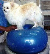
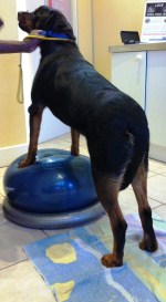
The best way to assure your dog is on the right track after his cranial cruciate ligament repair surgery is to incorporate a varied regimen of passive range of motion, balance exercises, and slow, controlled, leash walks. Range of motion exercises, also referred to as ROMs, will be demonstrated to you by our vet before you undertake them on your own, as you can do damage to the healing tissues in the knee with the wrong technique. Balance exercises can also be tricky for unskilled owners, and it is best that our qualified veterinary physical therapist perform balances exercises with your dog; think placing your dogs belly on a ball while helping him to bear weight and flex the knee.
Hydrotherapy/Swimming is an excellent non-weight bearing activity, but only after the incision site itself has had time to heal in the case of the lateral suture technique, and subsequent to the five week X-rays in the case of MMP surgery. All therapies should always be cleared through your veterinary surgeon, prior to their implementation and hydrotherapy should only be performed by a certified canine hydrotherapist. Hydrotherapy can be a great way to help them regain lost muscle mass while working on ROM exercises in a zero resistance environment (decreasing the likelihood of injury).
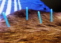
The use of animal acupuncture in a dog recovering from CCL repair surgery may help alleviate post op discomfort, which can help your dog to heal more quickly from surgery. Acupuncture can be used in conjunction with physical rehabilitation and should only be performed by a vet trained in acupuncture.
Our vet will provide you with a home physical rehabilitation plan which will include helpful post-op suggestions for your home environment. Examples are: securing rugs, adding rubber backed throw rugs for ease of walking on slippery surfaces, adding ramps to outside stairs and utilizing products for the paws to reduce falls.
The long term prognosis for animals undergoing surgical CCL repair is good, with clinical reports of improvement in 85-90% of the cases. Unfortunately, degenerative joint disease or osteoarthritis progresses regardless of treatment. Long term outcome includes a decrease in activity over time, an increasing level of disability, an adverse response to cold weather, and stiffness after inactivity related to progressive degenerative joint. Weight loss, an exercise regime of daily moderate activity, and the use of joint supporting supplements like glucosamine and chondroitin can help to improve these adverse clinical symptoms.
Read more about physical rehabilitation for pets.
Read more about acupuncture, laser therapy and stem cell therapy for pets.
back to Pet Info
back to Services
back to Home
What is Cranial Cruciate Ligament Rupture?
The canine knee (stifle) is a complicated joint. Two major bones meet in this joint, the femur (thigh bone) and the tibia (shin bone). These are joined by the patella (knee cap) and fabellae (very small bones found at the back of the knee). The joint is held together and stabilised by various ligaments. These ligaments make sure that the knee can only bend in ways that it should. There are two cruciate ligaments, the cranial (towards the head) and the caudal (towards the tail). Besides these ligaments there are tendons that attach the bones to muscles and so enable movement of the joint, and menisci which act as shock absorbing cushions.
Within this complicated joint the cranial cruciate ligament has the job of preventing the tibia from moving forward under the femur. When it ruptures, partially or completely, the tibia is able to move forward. This abnormal function results in varying degrees of lameness. The ruptured ligament makes the joint unstable, causes pain, and damage to the joint (for example, tearing of the shock absorbing cushions). If left untreated your dog's lameness may appear to improve in the first week or so, but you will note that the joint looks swollen and lack of treatment simply means that the abnormal movement caused by the ruptured ligament will continue and further degeneration of the joint will occur. In the long term osteoarthritis will develop even in cases that are diagnosed and treated in the acute stages of this disease, but the progression is quicker in chronic cases.
Rarely traumatic, cranial cruciate ligament rupture is more usually the result of a disease process extending over months or years. It is caused by a combination of several factors including degeneration of the ligament, obesity, poor physical condition, conformation, and breed. Many dogs who suffer from cranial cruciate ligament (CCL) rupture in one knee will go on to develop a similar problem in their other knee, and partial tearing of this ligament frequently progresses to a full rupture over time.
What are the signs and symptoms of Cranial Cruciate Ligament Rupture?
The degree of lameness varies from cases to case, but with complete CCL rupture it is likely that you will observe your dog becoming suddenly lame. However, given that many dogs suffer a partial tear and only progress to a full rupture over time it is important to be alert to more subtle signs including your dog's inability to sit square and preference for putting his legs out to the side when sitting down. Other signs to look out for include:
difficulty rising
difficulty jumping into the car
decreased activity level
muscle atrophy (decreased muscle mass in the affected leg)
decreased range of motion of the knee joint
a popping noise (which may indicate a meniscal tear)
swelling on the inside of the joint (fibrosis or scar tissue)
lameness
In chronic cases where the partial tear or rupture occurred some time ago the signs and symptoms will be akin to those seen with osteoarthritis:
decreased activity
stiffness
unwillingness to play
pain
In addition to taking a history from you, your veterinarian will manipulate the joint in order to test whether and to what extent the tibia is able to move forward under the femur. She may also take x-rays to confirm the presence of fluid in the joint, the progression of osteoarthritis, to aid in planning surgical treatment, and to rule out any other conditions that may be present. These x-rays will not enable your veterinarian to establish the extent of damage to the CCL or to the menisci. This can only be established through surgical investigation.
What treatments are available?
Surgical and non-surgical (also known as conservative) treatments are available, and your veterinarian will assist you in determining which is most appropriate for your dog. The most appropriate option for your pet will depend on your pets activity level, size, age, and conformation, the degree of knee instability, and other factors, such as the progression of osteoarthritis.
Surgical treatments are generally preferable when dealing with CCL rupture since they provide a means of permanently addressing the instability of the joint and any damage to the menisci. The surgical options available for controlling the joint's instability are varied and will depend on whether the surgery is carried out by a general practitioner or a specialist.
Specialist surgeons may choose to use procedures such as the Tibial Plateau Levelling Osteotomy (TPLO), Tibial Tuberosity Advancement (TTA) or Modified Maquet Procedure (MMP), which seek to address and control the instability caused by the ruptured CCL by changing the biomechanics of the joint. These techniques involve making a cut in the tibia. These procedures have demonstrated high degrees of success in enabling dogs to return to normal function after CCL rupture with a low level of complications.
The surgical techniques which involve cutting the tibia, TPLO, TTA, and MMP, have superior results with regard to limb function and the progression of osteoarthritis compared to traditional suture techniques, and are often favoured in cases involving young, large breed dogs. The disadvantage of such techniques is related to the cut made in the bone which requires time to heal, and any problem with this healing or with an implant may require further surgery.
Alternatives to these bone cutting surgeries include traditional suture techniques, particularly extra-articular (outside the joint) procedures. The most common is extra-articular (lateral) suture stabilization. These techniques use strong suture material to mimic the CCL and prevent the tibia moving forward under the femur. The suture material allows normal function whilst preventing abnormal function and gives time for scar tissue to form and assume the role of stabilising the joint. The disadvantages of such techniques include rupture of the suture material and the progression of osteoarthritis, whilst the advantages include lower costs and lack of any bone cutting (and so the removal of complications associated with cutting the bone).
Here at Southern Cross Veterinary Clinic we are able to offer both advanced cruciate ligament repair surgeries and lateral suture techniques. For all dogs over 15kg, TPLO, TTA and MMP are the preferred surgical techniques.
Non-surgical treatments generally involve pain management, restriction of activity, weight loss/control, therapeutic exercises and physical rehabilitation techniques to promote maintenance of function and management of osteoarthritis, and the use of custom knee braces or orthotics. These non-surgical treatments manage the patient's condition without addressing directly the stabilising role previously performed by the CCL. Nevertheless, physical rehabilitation therapy plays a crucial role in the post-operative recovery of surgical patients. It is highly recommended that such treatment begins immediately post-surgery.
How will treatment help this condition?
Prognosis for dogs treated surgically for CCL rupture is good, with clinical improvement being reported in 85-90% of cases. The surgical techniques address the instability of the joint either by changing the way it works by altering its biomechanics, or by mimicking the role performed by the intact CCL. Consequently, surgical intervention promotes normal function whilst preventing abnormal function. This enables as near a return to normal use of the knee as possible. Additionally, certain surgical techniques surgery allow for damage to the menisci to be assessed and addressed, something which is not possible with non-surgical treatments. This not only removes a source of pain and discomfort, but also an obstacle to full recovery, since dogs with torn menisci which are not removed will not return to near normal function of the knee. Unfortunately, osteoarthritis progresses regardless of the treatment option chosen and so management of this condition (read more about osteoarthritis) is necessary for the rest of your dog's life.
Post-surgical care is the highest priority in the treatment of CCL rupture. This will involve management of pain, restriction of exercise/activity, and physical rehabilitation techniques. In addition to medical management of pain, other techniques such as acupuncture, TENS (transcutaneous electrical stimulation), laser therapy, and cold therapy may be used to great effect to promote healing, relieve pain, and reduce inflammation. It can take dogs 6-7 months to walk normally following cruciate surgery, though physical rehabilitation can help to speed up this recovery and limit any complications.
Dogs can lose a third of the thigh muscle mass in their affected leg within 2 weeks of cruciate repair surgery and this muscle loss can continue for up to 5 weeks post-surgery. Therefore it is very important that physical rehabilitation begins immediately post-surgery and continues for at least 5 weeks. Initially physical rehabilitation helps to get your dog weight bearing as soon as possible after the surgery and so help to prevent muscle wasting. At Southern Cross in hospital rehabilitation begins even before your dog leaves theatre using techniques such as range of motion exercises and the application of ice packs. Our goal is to have your dog taking very good weight on the affected leg by the time of discharge which is about 2-3 days after the surgery depending on the surgical technique used.
At home you will need to keep your pet confined and make sure that he doesn't get the opportunity to run around, jump, or climb stairs or onto furniture. If your dog has had TPLO, TTA or MMP surgery it is essential that he is kept on leash until advised otherwise by the veterinarian. However, you will be given a home exercise plan by our physical rehabilitation trained veterinarian so that you can participate in your pet's recovery. This home exercise plan will build on the inpatient care provided immediately after surgery, and will complement the outpatient rehabilitation programme applicable to type of surgery your dog has undergone. Such a programme will include such techniques as passive range of motion, slow lead walking, massage, therapeutic exercises, hydrotherapy, and the use of cold and heat therapies. The aim of physical rehabilitation post-surgery will be to promote a return to as normal function as possible as quickly as possible by addressing muscle atrophy, joint use and function, gait, reduction of inflammation and pain relief.
Physical Rehabilitation/Therapy after Cruciate Surgery
The benefits of rehabilitation are well documented. Several university studies have now demonstrated the positive benefits to physical rehabilitation following cranial cruciate ligament (CCL) repair. Rehabilitation has been documented to improve muscle mass and attenuate muscle atrophy that occurs following surgery. Studies done by the American College of Veterinary Surgeons have shown that dogs beginning rehabilitative therapy within 48 hours of the procedure tend to heal faster and have greater range of motion than dogs without any physical therapy.
Without physical rehabilitation, dogs commonly lose 30% (or more) of their muscle mass within 2-3 weeks post-operatively. This loss of muscle increases the concussive forces through the joint and adds significantly to joint stress. 80% of joint stability is from the muscles and tendons so strong muscles mean more stable joints. Studies also demonstrate that rehabilitation speeds the return of both passive and active range of motion thereby increasing joint flexibility and adding to the overall speed of recovery and earlier weight bearing. Rehabilitation increases blood and lymph flow through the affected area, promotes early resolution of inflammation, prevents joint contractures, and reduces compensatory injury of other limbs. All of these factors reduce the incidence and progression of arthritis, decrease pain, lower complication rates, speed recovery and improve canine/ owner satisfaction.
The immediate post-op goal apart from reducing pain, inflammation and swelling is to have your dog weight-bearing as soon as possible after the surgery to prevent any muscle atrophy. Our goals will vary depending on the type of surgery. In the case of the lateral suture technique we aim to have your dog toe-touching within 24 hours of cruciate repair surgery and to have your dog taking good weight on the operated leg within 3-5 days following surgery. However, if your dog has undergone MMP surgery we aim to have your dog taking weight confidently within a day or two of surgery.
The best way to assure your dog is on the right track after his cranial cruciate ligament repair surgery is to incorporate a varied regimen of passive range of motion, balance exercises, and slow, controlled, leash walks. Range of motion exercises, also referred to as ROMs, will be demonstrated to you by our vet before you undertake them on your own, as you can do damage to the healing tissues in the knee with the wrong technique. Balance exercises can also be tricky for unskilled owners, and it is best that our qualified veterinary physical therapist perform balances exercises with your dog; think placing your dogs belly on a ball while helping him to bear weight and flex the knee.
Hydrotherapy/Swimming is an excellent non-weight bearing activity, but only after the incision site itself has had time to heal in the case of the lateral suture technique, and subsequent to the five week X-rays in the case of MMP surgery. All therapies should always be cleared through your veterinary surgeon, prior to their implementation and hydrotherapy should only be performed by a certified canine hydrotherapist. Hydrotherapy can be a great way to help them regain lost muscle mass while working on ROM exercises in a zero resistance environment (decreasing the likelihood of injury).
The use of animal acupuncture in a dog recovering from CCL repair surgery may help alleviate post op discomfort, which can help your dog to heal more quickly from surgery. Acupuncture can be used in conjunction with physical rehabilitation and should only be performed by a vet trained in acupuncture.
Our vet will provide you with a home physical rehabilitation plan which will include helpful post-op suggestions for your home environment. Examples are: securing rugs, adding rubber backed throw rugs for ease of walking on slippery surfaces, adding ramps to outside stairs and utilizing products for the paws to reduce falls.
The long term prognosis for animals undergoing surgical CCL repair is good, with clinical reports of improvement in 85-90% of the cases. Unfortunately, degenerative joint disease or osteoarthritis progresses regardless of treatment. Long term outcome includes a decrease in activity over time, an increasing level of disability, an adverse response to cold weather, and stiffness after inactivity related to progressive degenerative joint. Weight loss, an exercise regime of daily moderate activity, and the use of joint supporting supplements like glucosamine and chondroitin can help to improve these adverse clinical symptoms.
Read more about physical rehabilitation for pets.
Read more about acupuncture, laser therapy and stem cell therapy for pets.
back to Pet Info
back to Services
back to Home